
Computer-assisted Image-guided Interventions
Head: Prof. Dr. Andreas Melzer
ICCAS researches new technologies for computer-assisted image-guided procedures. This theme is a logical development of the traditional ICCAS research on computer-assisted surgery towards less invasive image-guided surgery and interventions.
Teams
Bio Signal Processing
Research focus: Computer-assisted Image-guided Interventions
Team lead: Michael Unger
Members: Reinhard Fuchs, Sven Martin, Mohamed Awad, Martin Hertel
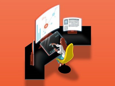
The team focuses on the use of sensors and technologies to improve medical practice – in diagnostics, treatment and training. One focus is non-invasive neurological mapping: neuronal activity is recorded precisely through the measurement of muscle activity, visualized and processed so that it can be seamlessly integrated into clinical workflows.
Further projects improve surgical training through data-based assistance systems that analyze training progress and provide targeted feedback, and create realistic simulation models with integrated sensors that reproduce anatomical conditions and biomechanical reactions in detail.
Projects
-
CortexMap Development of a novel navigated transcranial magnetic stimulation system for efficient planning and evaluation of neurosurgical procedures, as well as optimal integration into the surgical workflow
Navigated transcranial magnetic stimulation system for non-invasive mapping of the motor cortex in patients with brain tumors – with multi-channel EMG, automatic adjustment of stimulation intensity and analysis for surgical planning.
-
EyeHearU Multimodal acquisition, simulation and audiovisual enhancement for the individual training of basic functional laparoscopic skills
Training assistance system that records multimodal data from laparoscopic exercises and predicts training progress; two studies with students have been evaluated.
-
FaszioTrain Development of a sensor-based medical training model for fasciotomies
Sensor-based training model for fasciotomies of the lower leg that realistically reproduces swelling and pressure on the nerve pathways.
-
ReaFBS Feedback system for reducing brain damage during cardiopulmonary resuscitation
Feedback system that measures arterial blood flow during resuscitation using Doppler ultrasound – usable by laypersons without additional training; a mobile device with automated vessel location is in development.
-
NeoTapi Development of a non-invasive and contactless method for measuring physiological parameters for the detection and classification of apnea of prematurity and neonatal sepsis
Contactless measurement of respiration, heart rate and body temperature for detecting apnea of prematurity and neonatal sepsis.
Imaging Technologies
Research focus: Computer-assisted Image-guided Interventions
Team lead: Hannes Köhler
Members: Annekatrin Pfahl, Denis Gholami Bajestani, Julia Pretschner
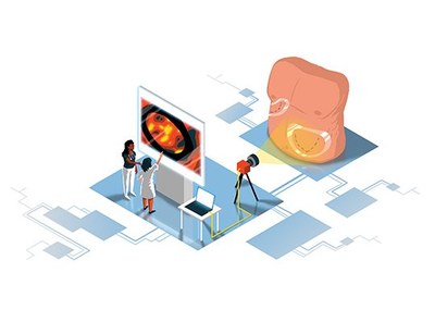
The team researches new imaging technologies and methods for non-invasive use in medicine. The focus is on optical procedures for perfusion imaging and tissue differentiation in the context of surgical interventions – from evaluation in the laboratory to the analysis of study data, in close cooperation with several clinics and industrial partners.
The team has many years of experience in spectral imaging, which can support the intraoperative assessment of tissue perfusion and the identification of tissue structures and tumor tissue. To support the transfer to clinical application, methods for the automatic classification and visualization of tissue are developed – using conventional machine learning as well as neural networks. The team supports clinical studies and supervises student theses in medicine, computer science and engineering.
Projects
-
 Klinische Relevanz von MSI und HSI Basic studies on the clinical relevance of multi- and hyperspectral imaging – industry-sponsored research by KARL STORZ SE & Co. KG
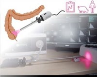
Basic studies on the clinical relevance of multi- and hyperspectral imaging (industry-sponsored research for KARL STORZ SE & Co. KG). In 2025 the first clinical study on nerve identification using laparoscopic HSI was published and an animal study on video-rate perfusion imaging was launched; a patent application was filed and a further patent granted.
Interdisciplinary Robotics
Research focus: Computer-assisted Image-guided Interventions
Team lead: Johann Berger
Members: Albrecht Bloße, Manuel Rosenau, Julia Pretschner
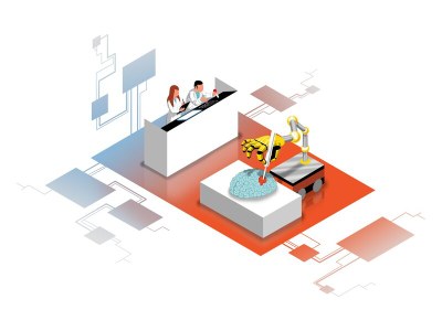
Since September 2024, the team has been working on the further development of medical robotic systems by integrating principles of soft and continuum robotics as well as networking and standardization. Robots made of flexible, soft materials are highly adaptable and safe when handling sensitive tissue; continuum robots do not require rigid joints and adapt precisely to complex geometries.
The aim is to make diagnostic and interventional robotic systems safer, more user-friendly and more efficient in order to facilitate their clinical application. The team develops soft end effectors such as gripping or guidance systems and realistic, patient-specific organ models – such as an actuated heart simulator – for planning, training and simulation. At the same time, it develops standardized descriptions of robotic systems and integrates them with other medical devices via IEEE 11073 SDC.
Projects
-
 SoKoRoMed Soft and Continuum Robotics for Medical Applications
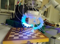
Soft and continuum robotics for medical applications: processes for producing patient- and application-specific soft end effectors. The reference use case is an actuated heart model as a training system for transcatheter aortic valve implantation (TAVI); first 3D-printed cardiac and vascular models and an ultrasound test bench for materials were developed and tested.
MR Technology
Research focus: Computer-assisted Image-guided Interventions
Members: Andreas Eger, Till Handel
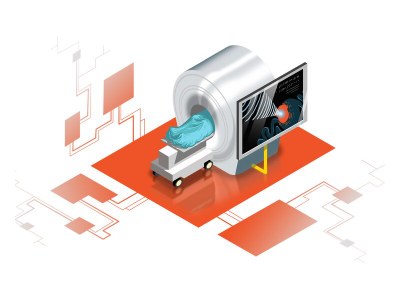
The project group deals with the development of MR-supported therapy procedures and the transfer of minimally invasive interventions, which are currently primarily performed using X-ray imaging, to the MR environment. The advantages are improved soft tissue contrast and the absence of iodine-containing contrast agents and ionizing radiation – which benefits patients as well as the hospital staff involved. The development of MRI-compatible instruments and devices is a central objective.
Our main focus is on therapy procedures using focused ultrasound (FUS) in MRI: the sound waves can be bundled in a focus approximately the size of a pine nut and used therapeutically without affecting the surrounding tissue. We are particularly interested in low-energy FUS for neuromodulation: it can activate or inhibit ion channels of neurons and thus non-invasively modulate deep brain areas – for example to treat stroke, addiction or obsessive-compulsive disorder.
Projects
-
 3MP-FUS Multimodality Multi-purpose Multi-platform Focused Ultrasound – Neuromodulation in rare neuropsychiatric disorders with focused ultrasound
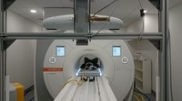
Multi-purpose, multi-parameter, multi-platform FUS system for transcranial neuromodulation in rare neuropsychiatric disorders. In 2025 the neuro-navigated system was integrated with Localite into the 1.5 T MRI (automatic slice orientation along the acoustic lobe) and transferred to the 3.0 T MRI at MPI CBS, where a neuromodulation pilot on volunteers is being prepared.
-
MRgFUS Non-invasive Magnetic Resonance Guided Focused Ultrasound in the treatment of uterine fibroids
Non-invasive treatment of symptomatic uterine fibroids with MR-guided focused ultrasound – with real-time temperature mapping and a very low complication rate.
-
Imaging Robotics Development of an integrated robotic system for the application in focused ultrasound interventions
Modular control software for robotic arms (KUKA LBR iiwa/Med) in image-guided interventions, connected via IEEE 11073 SDC; winner of the Best Design Award at the Hamlyn Symposium on Medical Robotics 2023.